Understanding the Mechanisms Behind Targeted Brain Therapy
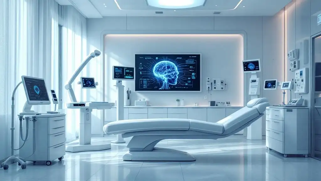
Transcranial Magnetic Stimulation (TMS) has revolutionized neurotherapy by offering a noninvasive method to modulate specific brain regions. Its ability to target neural circuits with precision hinges on scientific principles, advanced technology, and neuroimaging techniques. This article explores how TMS achieves targeted stimulation, the underlying mechanisms, and the integration of cutting-edge research for personalized treatment.
Fundamental Principles of TMS Targeting
How does TMS stimulate neurons?
TMS works by applying rapidly changing magnetic fields to the brain, usually lasting around 200 microseconds. These magnetic fields induce small electrical currents within the brain tissue through electromagnetic induction. The electrical currents then depolarize neurons by interacting with their axons and neural circuits, especially when the coil's magnetic field is accurately aligned perpendicular to neuronal structures. This activation can either excite or inhibit neural activity, depending on the stimulation parameters.
What is the scope of the stimulation area?
The area affected by TMS generally covers several square centimeters. Its size depends on multiple factors including the shape of the coil, the intensity of the stimulus, and the brain's electrical properties. For example, a figure-of-eight coil produces a more localized and focused stimulation, suitable for targeting specific cortical regions. Conversely, deep TMS coils can stimulate wider and deeper brain regions, enabling the treatment of more extensive neural networks.
Parameters influencing stimulation
The effectiveness and focus of TMS depend on various parameters such as stimulus intensity, coil orientation, and the electrical properties of the cortex. The placement position, using mapping techniques like motor threshold assessment, helps optimize coil orientation for maximal neuronal activation. Additionally, the frequency of pulses (high-frequency for excitation, low-frequency for inhibition) influences whether neurons are activated or suppressed. Interestingly, the brain's current state, cortical excitability, and recent activity also play significant roles in determining the outcome of stimulation. This highlights the importance of considering brain state dependency to improve therapeutic results.
| Aspect | Influence | Additional Details |
|---|---|---|
| Magnetic field | Induces electrical currents | Focused by coil shape and placement |
| Stimulation area | Several cm² | Dependent on coil design and intensity |
| Neuronal response | Depolarization or inhibition | Based on pulse frequency and brain state |
| Brain tissue properties | Affect current spread | Electrical conductivity varies across cortex |
Understanding these principles helps tailor TMS treatments to individual patients, aiming for optimal stimulation of targeted neural circuits.
Factors Determining the Reach and Specificity of TMS
How does coil orientation affect neuronal activation?
The orientation of the TMS coil is crucial in determining which neurons are stimulated. Proper alignment, typically perpendicular to the neuronal fibers, enhances the likelihood of depolarizing target neurons effectively. For instance, a figure-of-eight coil, which offers higher focality, is positioned to maximize the induced electric field over specific superficial cortical areas.
Why is TMS mainly effective for superficial brain regions?
The magnetic fields generated by TMS decrease steeply with increasing distance from the coil. This rapid fall-off means that while superficial cortical layers are readily stimulated, deeper brain structures receive significantly less current. Although advanced coils designed for deep TMS can penetrate further, the fundamental limitation remains that magnetic strength diminishes rapidly, confining effective stimulation mostly to accessible cortical regions.
What factors limit the depth of TMS targeting?
Effective stimulation depth is restricted by how quickly the magnetic field strength drops as it moves away from the coil. To reach deeper brain areas, clinicians may use specific coil configurations or increase stimulus intensity. However, such approaches often sacrifice the focality of stimulation and can affect surrounding tissues, highlighting a trade-off between depth and precision.
| Factor | Effect on TMS Reach | Additional Details |
|---|---|---|
| Coil shape | Determines focality and depth | Figure-of-eight coils offer focused stimulation; others may reach deeper but less precisely |
| Stimulus intensity | Extends possible stimulation depth | Higher intensity can activate deeper regions but risks non-specific activation |
| Electrical properties of cortex | Influences how current spreads | Variations in cortical tissue conductivity affect stimulation spread |
Understanding these factors helps optimize TMS protocols for targeted brain intervention, balancing depth, focality, and safety.
Neural Circuits and Network Modulation by TMS

How does TMS influence neural circuits?
Transcranial Magnetic Stimulation (TMS) not only affects the neurons directly beneath the coil but also induces widespread changes in brain networks. When TMS applies a brief, high-intensity magnetic pulse, it causes local depolarization of neurons in superficial cortical regions like the dorsolateral prefrontal cortex (DLPFC). However, these local effects extend beyond the stimulation site due to the brain's interconnected networks.
Through neural pathways, TMS modulates activity in remote areas that are functionally connected to the target region, leading to alterations in both structural and functional connectivity. This network-level influence can promote neuroplasticity—strengthening or weakening connections—and induce long-lasting changes in brain activity patterns. Such modulation is particularly relevant in psychiatric conditions like depression, where disrupted network connectivity within limbic and cognitive circuits contributes to symptoms.
What is the significance of brain state in TMS effects?
The impact of TMS largely depends on the current state of the brain during stimulation. Factors such as ongoing oscillatory activity, cortical excitability, and recent neural activity history influence how neurons respond to magnetic pulses. For instance, neurons that are near their firing threshold are more responsive to stimulation, resulting in more pronounced effects.
Aligning TMS with optimal brain states—such as specific phases of neural oscillations—can enhance efficacy, making treatment more personalized and effective. This dynamic interplay suggests that brain state-dependent stimulation could optimize outcomes, especially in therapies targeting neuroplasticity and network reconfiguration. Despite this, many current clinical protocols overlook this factor, potentially limiting treatment success.
Overview of brain network effects and their importance
| Effect Type | Description | Impact on Treatment |
|---|---|---|
| Local Activation | Depolarization of neurons near the coil | Immediate neural response |
| Remote Network Modulation | Changes in connected regions via neural pathways | Long-term plasticity, mood regulation |
| Neuroplasticity | Structural and functional changes in circuits | Sustained symptom improvement |
| Brain State Dependency | Variations in brain response based on current activity | Personalized, optimized protocols |
Understanding and harnessing these mechanisms can lead to more effective TMS treatments by strategically targeting the brain's networks in appropriate states, ultimately improving therapeutic outcomes.
Target Regions in TMS Therapy for Psychiatric Conditions

Which brain regions are involved in TMS therapy?
TMS therapy targets specific areas of the brain that are involved in mood regulation and emotional processing. A primary target in depression treatment is the dorsolateral prefrontal cortex (DLPFC), especially on the left side. This region helps regulate emotions and decision-making. Other important areas include the medial prefrontal cortex (MPFC) and the anterior cingulate cortex (ACC), which play roles in managing emotions and cognitive control.
In addition to these cortical targets, TMS can influence deeper and interconnected subcortical regions, such as regions of the limbic system involved in emotions and mood regulation. The overall goal of stimulating these areas is to restore balance and improve symptoms of mental health conditions like depression and OCD.
Why is the left DLPFC commonly targeted for depression?
In depressive disorders, the activity in the left DLPFC often tends to be diminished. This area is vital for positive emotional processing and decision-making. By stimulating the left DLPFC with high-frequency magnetic pulses, TMS aims to enhance its activity, which can help lift the depressive symptoms.
The lateralization hypothesis supports targeting the left side because increasing activity here supports mood improvement, whereas suppressing overactivity in the right DLPFC can also be beneficial. This focused approach helps to modify neural circuits involved in depression and aids in emotional regulation.
More insights into TMS brain targets
Further research has expanded TMS applications to include stimulation of interconnected regions such as the subgenual cingulate and nucleus accumbens. These areas are part of extensive brain networks involved in mood regulation and are accessible via advanced coil designs and stimulation protocols.
Neuroimaging tools like fMRI and PET scans help identify optimal targets and monitor treatment effects, demonstrating that TMS influences not just localized brain regions but also broader neural connectivity networks. This neuroplasticity supports the idea that TMS can promote lasting changes in brain function, providing new avenues for treatment of various psychiatric conditions.
| Brain Regions | Function | Relevance to TMS | Notes |
|---|---|---|---|
| DLPFC (Left) | Mood regulation, decision-making | Main target in depression | High-frequency stimulation enhances activity |
| DLPFC (Right) | Overactivity in some cases of depression | Overactivity reduction can help | Low-frequency stimulation can be used |
| MCC and MPFC | Emotional processing, cognitive control | Additional targets | Devices like H7 coil specifically target these areas |
| Subcortical regions (e.g., limbic system) | Emotions, motivation | Can be influenced indirectly | Advanced coil designs expand reach |
This comprehensive approach to targeting brain regions supports the personalized and effective use of TMS in treating mood disorders, improving patients' emotional well-being and cognitive functions.
Neuroimaging Techniques Informing TMS Targeting
 Modern neuroimaging methods play a crucial role in enhancing the precision and effectiveness of transcranial magnetic stimulation (TMS). Techniques such as magnetic resonance imaging (MRI), functional MRI (fMRI), positron emission tomography (PET), and diffusion tensor imaging (DTI) provide detailed insights into brain structure and function.
MRI offers high-resolution images of the brain's anatomy, helping identify the exact location of target regions like the dorsolateral prefrontal cortex (DLPFC). These images enable clinicians to tailor the placement of the TMS coil to the individual’s unique brain shape.
Functional imaging techniques like fMRI and PET reveal activity patterns and metabolic processes within brain networks. For example, in depression, these methods can show reduced activity in the left DLPFC and overactivity in other areas, guiding targeted stimulation to restore balance.
Diffusion tensor imaging (DTI) maps white matter pathways, illustrating how different brain regions are structurally connected. This information supports the personalization of TMS by targeting not only specific spots but also interconnected networks involved in mood regulation.
How do neuroimaging studies guide TMS targeting?
Neuroimaging methods such as MRI, fMRI, PET, and DTI help identify functional and structural brain regions involved in disorders like depression. These techniques allow clinicians to localize optimal stimulation sites and assess connectivity patterns, leading to personalized treatment plans.
What role does functional connectivity play in targeting?
Functional connectivity analysis reveals how different brain areas communicate and respond to TMS. By understanding these networks, clinicians can choose targets that modulate entire circuits rather than isolated regions, enhancing therapeutic outcomes.
Exploring Additional Insights
| Imaging Modality | Main Use | Role in TMS | Additional Details |
|---|---|---|---|
| MRI | Structural imaging | Localize target regions | Provides detailed brain anatomy for precise coil placement |
| fMRI | Brain activity mapping | Identify hypo- or hyperactive areas | Guides stimulation of underactive regions associated with depression |
| PET | Metabolic activity | Assess brain function | Helps find dysfunctional circuits for targeted therapy |
| DTI | Brain connectivity pathways | Map white matter tracts | Facilitates targeting interconnected networks |
By integrating these neuroimaging techniques, clinicians can design more effective, individualized TMS treatments, ultimately improving patient outcomes through precise and network-informed stimulation.
Technical Approaches for Precision in TMS Application

How is neuronavigation used in TMS?
Neuronavigation systems play a crucial role in enhancing the accuracy of TMS treatments. These systems leverage detailed neuroimaging data, such as magnetic resonance imaging (MRI), to guide the precise placement of the coil over the targeted brain region. By integrating real-time tracking of the coil position with the patient's own brain anatomy, clinicians can ensure the magnetic pulses are delivered exactly where they are needed. This precision reduces variability in treatment delivery and can improve overall therapeutic outcomes, especially when targeting specific cortical areas like the dorsolateral prefrontal cortex (DLPFC).
What role do computational models play?
Computational modeling is an essential tool for optimizing TMS parameters. By simulating electric field distribution within the brain, researchers can predict how different coil configurations, orientations, and stimulus intensities affect various brain regions. Finite element models (FEM), for example, can take into account the electrical properties of cortical tissue to enhance the precision and efficacy of stimulation. These models help in designing personalized treatment protocols, enabling clinicians to tailor stimulation to the specific anatomy and needs of individual patients.
How does multimodal imaging enhance targeting?
The integration of multiple neuroimaging techniques further refines TMS targeting strategies. Combining functional MRI (fMRI), EEG, and other modalities provides a comprehensive view of brain activity and network connectivity. This approach allows for identification of the most relevant neural circuits involved in a mental health condition, such as depression or OCD. By understanding how different regions interact within individual brains, clinicians can customize stimulation protocols to modulate specific networks, potentially improving treatment response. Such multimodal data enrich the understanding of brain states and guide the timing and location of TMS pulses.
| Approach | Main Technique | Benefits | Additional Details |
|---|---|---|---|
| Neuronavigation | MRI-guided real-time tracking | Precise coil placement, reduced variability | Guided by individual neuroanatomy, improves targeting accuracy |
| Computational modeling | Finite element analysis | Optimized stimulus parameters, personalized treatment | Simulates electric fields, predicts stimulation outcomes |
| Multimodal imaging | MRI, EEG integration | Network-focused, tailored protocols | Addresses individual neuroarchitecture for better efficacy |
From Physics to Clinical Practice: Administering TMS

How is TMS administered for targeted therapy?
Clinicians deliver TMS by placing an electromagnetic coil against the patient's scalp, over the specific brain region targeted for treatment. The precise placement of the coil is often guided by neuroimaging techniques like MRI, which helps map individual brain anatomy. During sessions, pulses are issued with carefully controlled parameters, including frequency and intensity, to modulate neural activity effectively.
This method allows for noninvasive stimulation, primarily affecting superficial cortical regions. The goal is to activate or inhibit specific neuronal populations to influence brain circuits implicated in depression, anxiety, or other conditions.
What ensures accurate coil placement?
Coil positioning relies on advanced localization techniques. Neuronavigation systems integrate individual MRI data, providing real-time guidance to align the coil precisely over the target area. Additionally, measurements such as the motor threshold—elicited by stimulating the motor cortex and observing muscle responses—help define the appropriate stimulation intensity. These measures optimize the accuracy and consistency of stimulation across sessions.
Tracking and adjusting the coil placement to match the targeted brain region improves treatment efficacy, especially when stimulating deeper or specific subregions like the dorsolateral prefrontal cortex or medial prefrontal areas.
How are stimulation protocols personalized?
Personalization of TMS protocols hinges on neuroimaging findings and individual brain characteristics. MRI and other imaging modalities help identify the exact sites for stimulation based on the patient’s unique anatomy and pathology.
Protocols are also tailored considering the disorder’s nature. For depression, high-frequency stimulation generally aims to excite underactive areas like the left DLPFC, whereas low-frequency protocols might suppress overactive regions. Adjustments in pulse frequency, intensity, and session duration are made according to the patient's response and tolerance.
Emerging approaches include multi-target stimulation, where several interconnected regions are stimulated sequentially or simultaneously, potentially enhancing outcomes. This personalization process is crucial to increase the effectiveness of TMS therapy and optimize patient-specific brain modulation.
The Future of TMS: Enhancing Specificity and Effectiveness
 Emerging approaches in transcranial magnetic stimulation (TMS) focus on increasing its precision and expanding its capabilities for treating complex brain disorders. One major development is the adoption of deep TMS and multi-target strategies. Deep TMS uses specialized coils designed to stimulate deeper and broader regions of the brain, reaching areas involved in mood regulation and neuroplasticity that are inaccessible with standard TMS. Multi-target stimulation involves engaging several interconnected regions simultaneously, such as the dorsolateral prefrontal cortex (DLPFC), subgenual cingulate, and hippocampus, to produce more comprehensive therapeutic effects.
Advances in neurotechnology are also poised to enhance TMS outcomes. Combining TMS with neurofeedback or brain-computer interfaces (BCIs) allows for real-time monitoring of brain activity. This integration enables the adjustment of stimulation parameters on-the-fly, ensuring that neural modulation is accurately targeted during therapy sessions. Such adaptive approaches can improve treatment efficacy by responding to the brain's current state, rather than applying uniform stimulation protocols.
Looking ahead, personalization of TMS treatments is set to become more sophisticated. Future protocols are expected to tailor stimulation to each individual's unique brain connectivity profile and dynamic neural state. Using neuroimaging data (like MRI or EEG), clinicians can identify optimal stimulation sites and adjust pulse parameters to maximize therapeutic benefits. Adaptive, real-time algorithms could modulate stimulation based on ongoing neural feedback, allowing for truly individualized interventions that evolve with the patient's progress.
| Innovation Approach | Focus Area | Potential Benefits |
|---|---|---|
| Deep TMS coils | Stimulating deeper brain regions | Reaching circuits involved in depression and neuroplasticity |
| Multi-target stimulation | Engaging interconnected regions | More effective symptom relief and network modulation |
| Neurofeedback & BCI integration | Real-time brain activity monitoring | Increased precision and personalized adjustments |
| Adaptive protocols | Dynamic, patient-specific treatment | Improved response rates and reduced side effects |
As research advances, the horizon for TMS promises more targeted, adaptable, and effective treatments for neurological and psychiatric conditions, underpinning a new era of neurotherapeutic precision.
Transforming Neuroscience and Psychiatry
Understanding how TMS targets specific brain regions requires an appreciation of its complex scientific basis, technological advancements, and integration with neuroimaging. As research continues to refine its precision and personalization, TMS stands poised to offer increasingly effective therapies for mental health and neurological conditions, making brain circuit modulation a cornerstone of future neuropsychiatric treatment.
References
- How does transcranial magnetic stimulation modify ...
- Target Engagement and Brain State Dependence of ...
- Transcranial magnetic stimulation
- TMS Treatment Targets Specific Areas of the Brain
- Effects of repetitive transcranial magnetic stimulation and ...
- Transcranial Magnetic Stimulation (TMS) for Depression, ...
- Unlocking TMS: What Part of the Brain does TMS Stimulate?